Jae-Byum Chang LabMolecular Imaging and Neurodevices Lab @ KAIST

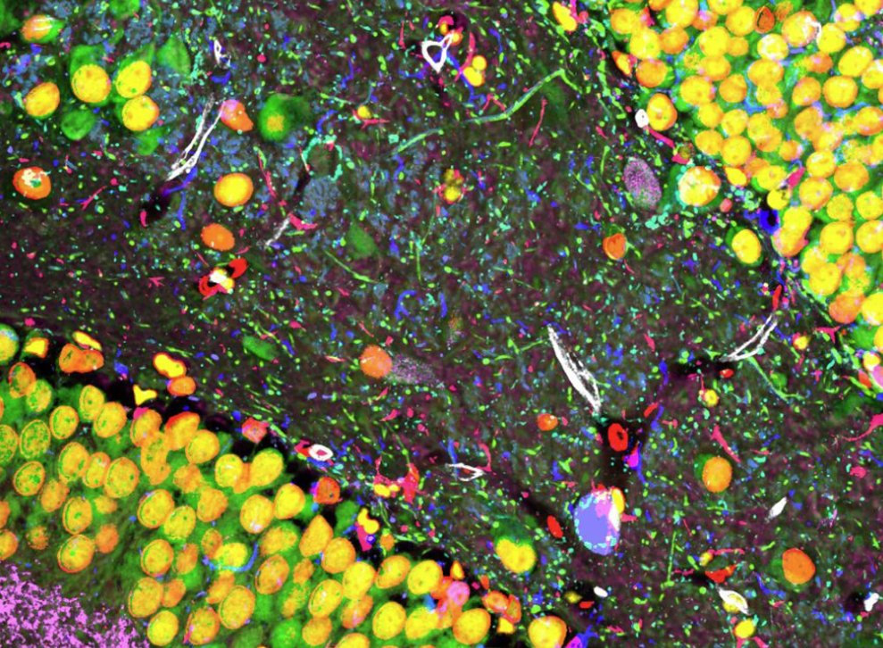
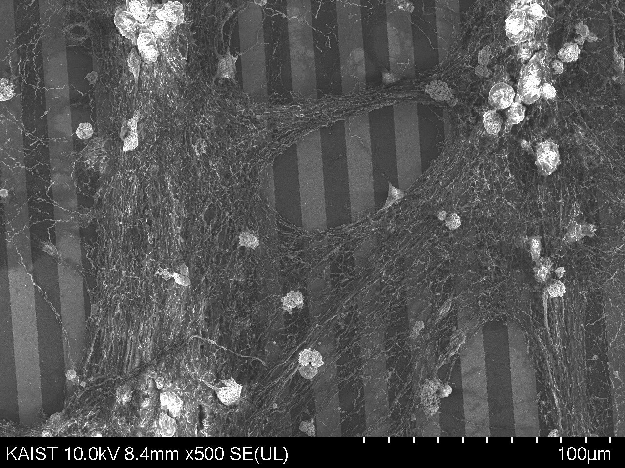
We treat tissue imaging as a precision engineering system — integrating novel chemistry, automated processes, AI-based analysis, and nanofabrication to produce large-scale, quantitative spatial biology data. Our goal is to map the molecular architecture of cancer and the brain at nanoscale resolution, and to translate these biological blueprints into next-generation semiconductor devices.

Spatial Proteomics Expansion Microscopy Biomaterial Synthesis AI-driven Analysis KAIST
Chang Lab research image 1 Chang Lab research image 2 Chang Lab research image 3

News

2026년 4월 (2026년 4-7월) 박사후연구원(Post-doc) 모집우리 연구실에서 박사 후 연구원을 모집합니다. 모집중 자세히 보기 ▼

우리 연구실에서는 아래와 같은 분을 박사후연구원으로 찾고 있습니다.

• Microfluidics, robotics, instrumentation, automation 등을 전공하였으며, 이를 최신 분자 이미징 기술에 접목해보고 싶은 분
• AI, machine learning, data science, computational imaging 전공자 중, 대량의 정밀 분자 데이터를 직접 얻고, 이를 분석 가공하여 새로운 생물학적 혹은 의학적 발견을 해보고 싶은 분
• 암, 신경과학, 세포생물학 등 생물학 연구를 수행해오신 분 중, multi-omics 기술 개발 및 적용에 관심이 많은 분
• 기타 기계, 전자, 전산, 화공, 재료, 생물, 의공학 등을 전공한 분 중, 새로운 이미징 기술의 개발, 이미지 획득, AI 분석, 생물학적 및 의학적 적용에 관심이 있는 분

관심 있는 분은 jbchang03@kaist.ac.kr로 CV와 간단한 소개를 보내주시기 바랍니다.
2026년 4월 (2026년 4-7월) 2026년 가을학기 및 2027년 봄학기 대학원생 모집KAIST 신소재공학과 또는 생명과학과 대학원으로 입학하는 학생을 모집합니다. 모집중 접기 ▲

우리 연구실은 첨단 분자 이미징과 차세대 neurodevice를 연구합니다.

우리는 expansion microscopy (ExM), multiplexed tissue imaging, imaging chemistry, hydrogel engineering, process automation, quantitative imaging을 포함한 통합 이미징 파이프라인을 구축하고, 이를 통해 복잡한 생체 조직에서 정확하고 재현성 높은 spatial biology 데이터를 생산합니다. 동시에, 실제 neuronal architecture를 기반으로 새로운 bioelectronic device 및 차세대 neuromorphic hardware를 개발합니다.

즉, 우리 연구실은 이미지를 단순히 얻는 데서 끝나지 않고,
샘플 준비–화학 처리–이미징–정렬/분석–정량화의 전 과정을 최적화하여 대규모 정밀 데이터를 만들고, 나아가 생물학적 구조를 새로운 기능성 소자와 하드웨어로 연결하는 연구를 수행합니다.

주요 연구 분야
• 공간단백체(spatial proteomics) 및 multiplexed imaging 기반의 암·뇌 조직 분석
• Expansion microscopy를 위한 새로운 chemistry 및 hydrogel engineering
• Multiplexed protein imaging 및 차세대 분자 이미징 기술 개발
• 대용량 생체 이미지의 정렬, 정량 분석, 자동화, 표준화 파이프라인 개발
• AI 기반 공정 자동화 및 데이터 기반 생물학적/의학적 해석
• 실제 neuronal architecture를 이용한 bioelectronic device 및 neuromorphic device 개발

이런 학생을 찾습니다
• 생명과학/의생명 계열 학생 중 첨단 분자 이미징, spatial biology, multi-omics, 정량 생물학에 관심 있는 학생
• 신소재/반도체/전자/물리/화학 계열 학생 중 공정, 소자, 자동화, 데이터 분석, 차세대 하드웨어에 관심 있는 학생
• 실험과 분석을 함께 배우고 싶거나, biology와 engineering을 연결하는 새로운 연구를 해보고 싶은 학생

우리 연구실에서 배우는 것
• ExM, mIF, spatial proteomics 등 첨단 분자 이미징 기술
• 샘플 preparation, labeling chemistry, imaging chemistry, hydrogel engineering
• 대규모 이미지 정렬, 정량 분석, AI 기반 데이터 해석
• 공정 설계, 자동화, 재현성/표준화 기반의 연구 수행 방식
• biological architecture를 차세대 device/hardware로 연결하는 융합 연구

졸업생 진로: Johns Hopkins University 등 세계 유수 대학 박사후연구원 (박사 3명), 삼성전자·삼성SDI·SK Hynix 등 반도체 및 소재 기업 (박사 4명, 석사 1명), 바이오 벤처 및 벤처 투자사 (박사 1명, 석사 3명), MIT·UC Berkeley 대학원 진학 (학부연구생 2명)

관심 있는 학생은 jbchang03@kaist.ac.kr로 CV와 간단한 소개를 보내주시기 바랍니다.
2025년 4월 MIT News에 장재범 교수 연구 소개"A brief history of expansion microscopy" 언론 보도

MIT McGovern Institute가 expansion microscopy 탄생 10주년을 기념해 게재한 특집 기사에서, 장재범 교수(Boyden 교수 연구실 출신)가 KAIST에서 쥐 배아와 제브라피시 전체를 4배 확장 이미징하는 whole-body ExM을 개발했다고 소개되었습니다. 뇌에서 척수를 거쳐 최종 목표까지 신경을 매핑하는 것이 다음 목표라고 밝혔습니다.

→ MIT News 기사 보기
2025년 2월 Wiley Analytical Science에 Chang Lab 연구 소개"Seeing it all: the rise of entire-organism expansion microscopy" 언론 보도

국제 과학 매거진 Wiley Analytical Science에 장재범 교수팀의 whole-body ExM 연구(ACS Nano)가 소개되었습니다. 쥐 배아 전체를 4배 균일 확장하여 해부학적 구조와 단백질을 초고해상도로 이미징하는 데 성공했으며, 향후 더 큰 확장 배율과 신경 연결 지도 작성으로의 확장을 목표로 하고 있다고 밝혔습니다.

→ Wiley Analytical Science 기사 보기
Jan 2025신개념 생체형틀법 캠바이오(CamBio) 개발 — 장재범·정연식 교수 공동연구팀. KAIST 뉴스 →Publication
Feb 2024송창우 박사과정, KAIST 신소재공학과 최우수 학술논문상 수상.Award
Aug 2022항체를 활용한 신개념 생체 형틀법 최초 개발Advanced Materials 게재. KAIST 뉴스 →Publication
May 2022암·뇌졸중·치매 등 난치병 진단을 위한 멀티 바이오마커 동시 탐지 기술(PICASSO) 개발 — Nature Communications 게재. KAIST 뉴스 →Publication
View all news →

Research

01

Integrated Molecular Imaging Pipeline

End-to-end optimization of the full imaging workflow — from tissue preparation and labeling chemistry to microscopy, image registration, and quantitative analysis — to generate accurate, reproducible, high-dimensional spatial datasets from cancer and brain tissues.

Molecular imaging Multiplexed imaging Image registration Quantitative analysis
02

Imaging Chemistry and Expansion Engineering

Chemical and materials strategies that enhance imaging capability — including expansion microscopy, hydrogel engineering, and labeling chemistry — to improve structural fidelity, signal accessibility, and multiplexing performance across diverse tissues.

Expansion microscopy ExM hydrogel engineering Labeling chemistry Structural fidelity
03

Molecular Imaging and Neurodevices

Engineering neuronal architectures through surface patterning and molecular control, then linking structural imaging readouts to metallization, electrical characterization, and device behavior — toward new bioelectronic and neuromorphic platforms.

Neurodevices Neuronal patterning Bioelectronics Neuromorphic systems
View full research →

Selected Publications

2025

Nanoscale Resolution Imaging of Whole Mouse Embryos Using Expansion Microscopy

Jueun Sim+, Chan E Park+, In Cho+, et al. · ACS Nano

ACS Nano →
2017

Iterative expansion microscopy

J. Chang, F. Chen, Y. Yoon, et al. · Nature Methods

Key publication Nature Methods →
View all 58 publications →

Team

Prof. Jae-Byum Chang

Jae-Byum Chang

Principal Investigator

WL

Woonggi La

PhD Student

CK

Chan Kang

PhD Student

YY

Youngbin Yoon

PhD Student

HJ

Hoeyun Jung

PhD Student

NK

Nakhyun Kim

PhD Student

JK

Jinsu Kim

PhD Student

EK

Eunwoo Kim

PhD Student

View full team →

Join us

Graduate students

We are actively recruiting motivated MS/PhD students with backgrounds in biology, chemistry, physics, or engineering. Positions are available each semester.

Inquire →

Postdoctoral researchers

Candidates with expertise in imaging, soft matter, or single-cell biology are encouraged to reach out with a CV and brief statement of research interests.

Inquire →

Undergraduate internships

Motivated undergraduates can join through UROP or informal internship programs. Reach out early in the semester.

Inquire →

Contact

Department of Materials Science and Engineering
KAIST, Daejeon, South Korea

jbchang [at] kaist.ac.kr